

For most of our lives, our bodies quietly maintain their own architecture. Skin stays elastic. Bone rebuilds itself. Joints glide. We don’t think about any of it, because we don’t have to — it simply happens in the background, the way a well-run machine takes care of itself.
Then, somewhere in our forties, the maintenance schedule changes. Often before a single new wrinkle appears, we notice something harder to name: a stiffness that wasn’t there last year, skin that seems to hold its shape differently, an energy in our joints that feels less certain. If you’ve felt this and wondered whether you were imagining it, you weren’t. What you were noticing has a name, a mechanism, and a growing body of research behind it: structural aging — a specific, measurable process that is meaningfully different from simply getting older.
At Monna, this distinction is where we start every conversation about aging, because understanding what is actually changing beneath the surface is the first step toward protecting it.
The Four Pillars of Structural Health
We think of structural health as the combined integrity of four collagen-dependent tissue systems. Each one ages on its own timeline, but all four are built from the same underlying biology — which is part of why they tend to shift together, not separately.
Skin
The dermis — the layer beneath the visible epidermis — is roughly 70–80% type I and type III collagen by dry weight. This structural collagen matrix, produced by cells called fibroblasts, is what gives skin its firmness, elasticity, and ability to hold moisture. When dermal collagen thins, skin doesn’t just look older; it is structurally thinner, less resilient, and slower to bounce back from everyday stress.
Bone
Bone is often described as a mineral structure, but roughly 90% of its organic matrix is type I collagen, arranged in a scaffold that mineral crystals attach to.¹ This structural collagen framework gives bone its tensile strength — the ability to bend slightly under load before it snaps. Bone mineral density (BMD) gets most of the attention, but the collagen scaffolding beneath it is just as responsible for whether a bone resists fracture.
Joint & Cartilage
Cartilage cushions the ends of our bones and is largely made up of type II collagen and proteoglycans, maintained by cells called chondrocytes. Unlike skin or bone, cartilage has very limited blood supply and a slow, limited capacity to repair itself and that’s why joint changes during midlife can feel so much more permanent than changes we see in the mirror.
Connective Tissue (Tendon, Ligament, Fascia)
Tendons, ligaments, and fascia are collagen-rich structures that transmit force, stabilize our joints, and hold the body’s systems together. These tissues are easy to overlook in conversations about aging but they are just as collagen-dependent and just as hormonally sensitive as skin.
Structural Aging vs. Chronological Aging
Chronological age is simply a number on a calendar. Structural age is the actual condition of these four structural collagen systems (skin, bone, joint & cartilage, connective tissue), and the two can be significantly different. Research on skin has found that collagen content correlates more closely with time since menopause than with a woman’s chronological age — meaning two 52-year-olds, one two years postmenopausal and another one eight years postmenopausal, can have meaningfully different structural profiles despite being born in the same year.² This is why we find structural aging to be a more precise, and more actionable, framework than simply counting birthdays.
The Estrogen–Collagen Connection: The Hormonal Driver Behind Structural Aging
If you’re wondering what’s really driving all of this, here’s the honest answer: it isn’t primarily diet, sleep, exercise, or sun exposure, although each of those matters. It’s estrogen.
Estrogen receptors (ERα and ERβ) sit directly on the fibroblasts that build our skin collagen, the osteoblasts that build our bone, and the cells that maintain our cartilage and connective tissue.³
Laboratory studies show that estrogen actively stimulates type I collagen production in dermal fibroblasts while simultaneously suppressing the enzymes (matrix metalloproteinases) that break collagen down.⁴ Estrogen, in other words, isn’t a passive bystander in tissue health. It’s one of the primary signals telling our bodies to keep building and repairing their own structural scaffolding.
So when estrogen production declines during perimenopause, and then falls sharply after menopause, that signal weakens. Collagen synthesis slows. Collagen breakdown accelerates. And the four pillars of structural health — skin, bone, joint, and connective tissue — all lose ground at roughly the same time. This is why so many changes that feel unrelated — drier skin, achier joints, a lower bone density score, tendons that take longer to recover after exercise — often arrive around the same time. They aren’t a coincidence of timing. They share a common cause: estrogen decline.
Why Structural Aging Accelerates During Perimenopause and Menopause
The data here is specific, and it’s worth looking into. Studies that directly measure skin collagen have found that women lose approximately 30% of their skin collagen in the first five years following menopause, with continued losses of roughly 2.1% per year for at least a decade after that — a decline tied to time since menopause rather than chronological age alone.⁵ Bone follows a similar pattern: in a 12-month randomized controlled trial of postmenopausal women with age-related bone density decline, the placebo group continued to lose bone mineral density, while women receiving daily specific collagen peptides saw significant increases in both spinal and femoral neck BMD.⁶
This is the structural reality of the menopause transition, and it’s one many of us were never told: it isn’t one symptom, or even a cluster of symptoms. Menopause is a system-wide shift in how the body maintains its own architecture, set in motion by the loss of the hormone that had been quietly coordinating that maintenance for decades.
Why This Reframe Matters for Longevity
Most conversations about aging start at the surface — a new serum, a different cream, a concealer for under-eye circles. We’d rather start with a more useful question: what is actually happening to the tissue underneath, and what does the evidence say can support it?
That question is the foundation of how Monna approaches female aging — not as a cosmetic problem to conceal, but as a biological process, rooted in the estrogen–collagen connection, that we can understand, measure, and support with the right evidence-based daily rituals. Structural health was never about chasing youth. It’s about protecting the architecture that lets each of us move, feel, and live well — for as long as possible.
¹ Viguet-Carrin S, Garnero P, Delmas PD. The role of collagen in bone strength. Osteoporos Int. 2006;17(3):319–336.
² Bhat YJ, Bashir S, Qayoom S, et al. Managing menopausal skin changes: a narrative review of skin quality changes, their aesthetic impact, and the actual role of hormone replacement therapy in improvement. PMC. Available at: pmc.ncbi.nlm.nih.gov/articles/PMC12374573 (citing Brincat M, et al., observational and histologic data on collagen decline of 2.1% per postmenopausal year over 15 years).
³ Markiewicz M, Znoyko S, Stawski L, et al. A role for estrogen receptor alpha (ERα) and estrogen receptor beta (ERβ) in collagen biosynthesis in mouse skin. J Invest Dermatol. Available at: pmc.ncbi.nlm.nih.gov/articles/PMC3502697.
⁴ Stevenson S, Thornton J. Effect of estrogens on skin aging and the potential role of SERMs. Clin Interv Aging. 2007;2(3):283–297; see also: Beneficial regulation of type I collagen and matrix metalloproteinase-1 expression by estrogen, progesterone, and its combination in skin fibroblasts. GeroScience. Available at: pmc.ncbi.nlm.nih.gov/articles/PMC3456089.
⁵ Bhat YJ, Bashir S, Qayoom S, et al. Managing menopausal skin changes: a narrative review of skin quality changes, their aesthetic impact, and the actual role of hormone replacement therapy in improvement. PMC. Available at: pmc.ncbi.nlm.nih.gov/articles/PMC12374573 (citing Brincat M, et al., observational and histologic data on collagen decline of 2.1% per postmenopausal year over 15 years).
⁶ König D, Oesser S, Scharla S, Zdzieblik D, Gollhofer A. Specific collagen peptides improve bone mineral density and bone markers in postmenopausal women — a randomized controlled study. Nutrients. 2018;10(1):97. doi:10.3390/nu10010097.
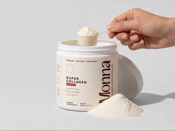
Super Collagen
Backed by 3 patented bioactive collagen peptides — built for perimenopause and menopause.